bone grafting
The ridge of your jaw needs adequate width and height of bone for long term success of your dental implant.
Bone loss is a well-documented result of dental extraction. The ridge loses bone volume in both height and width. This bone loss can compromise the ability to place an implant of ideal size in the ideal position to restore a tooth.
A common procedure is bone grafting a site to help restore volume of bone. There are different materials used for different situations encountered.
Bone grafting can be done at time of tooth extraction to help preserve the ridge. Roughly 50% of the ridge width is lost in the first year after extraction, and much of this occurs in the first few months. There is also a vertical loss of bone. Grafting at time of extraction is done to reduce bone loss. The purpose of maintaining or creating adequate ridge height and width is to allow an appropriate sized implant to replace the missing tooth in ideal position.

An upper premolar was extracted and no bone graft performed. The ridge atrophied as expected. Patient was interested in an implant. CT scan and virtual planning shows lack of bony support for an implant. A bone grafting procedure was performed and after a healing period the ridge had adequate volume to place an implant.

A patient had an upper premolar extracted at a dental office and presented for an implant. No bone grafting had been performed. 3D imaging and treatment planning software can identify the ideal implant position in the existing extraction site. The left image shows that an implant would not have adequate bony coverage for long term stability. The right image is after a bone augmentation procedure was completed to adequately cover and support a dental implant.

Molar tooth with root canal therapy with persistent infection. The darkness around the root is active infection causing bone loss. Tooth was extracted and bone grafted. After a healing period the augmented bone ridge had healed and was ready for an implant.
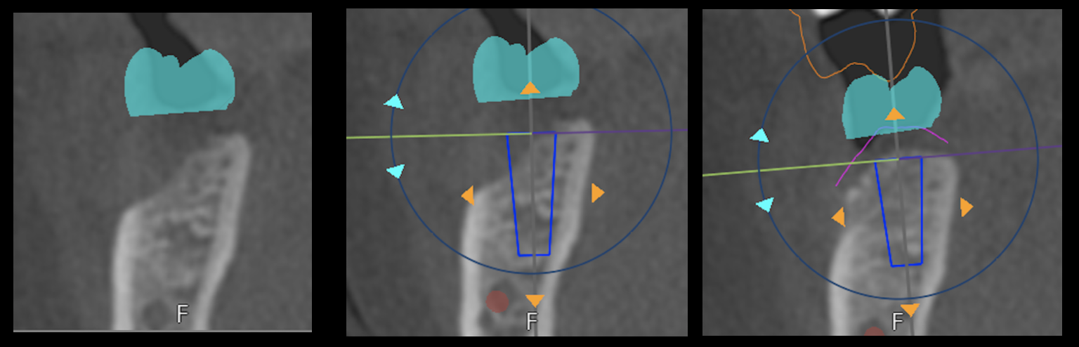
A tooth was extracted and no graft was placed. The left and center pictures are pre-graft. A CT scan was imported into treatment planning software with a virtual implant placed in ideal position. This shows a lack of bone to support the implant. A bone graft was placed to augment the ridge. Far right picture is of a CT scan shows adequate bony support for an implant. This is a great example of two things: 1. How bone loss occurs after a tooth extraction and 2. how a bone graft can rebuild a resorbed ridge to allow placement of an implant.

The left images are of a molar site where a tooth had been extracted at an outside office. No bone graft was performed. Patient presented about a year after the extraction to inquire about getting a dental implant. Cone beam CT scan was taken with planning software to show an implant and crown in an ideal position. This workup shows both the vertical and horizontal lack of bone. A bone graft was performed. Image on the right is the same site after the bone graft had healed. Adequate height and width of bone are available to place an implant in ideal position. Note: the red dot is the nerve, and planning software helps safely identify the nerve and plan and place the implant safely away from it.